(III) Bacterial cell structure
(Unit III ) Bacterial cell structure:
• Ultrastructure of Bacteria-
1.Cell wall (Gram Positive and Gramnegative),
2.Cell Membrane,
3.Capsule,
4.Flagella,
5.Pili,
6.slime layer,
7.Ribosome,
8. Nucleoid,
9.Mesosomes,
10.Endospore,
11.Cell inclusions (Gas vesicles, carboxysomes,magnetosomes, PHB granules, Glycogen bodies, metachromatic granules)
Ultrastructure of Bacteria-
1. Cell wall (Gram Positive and Gram negative)
Cell Wall:
Just outside the plasma membrane, a complex semi-rigid structure that determines the shapes of bacteria is called a cell wall.
The cell wall is an essential structure, as most bacteria cannot live without it.
Exceptionally Mycoplasma species and several archaea do not have cell wall.
Although presence of cell wall is an unique characteristics of bacteria. two kinds of cell walls are detected in bacteria -Gram-positive and Gram-negative . These categories are based on differential staining response due to difference in chemical composition of cell wall.
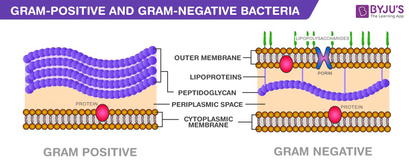
Gram-positive and gram-negative cells do share one thing in common that is unique to bacteria and it is the presence of peptidoglycan .
Structure of Gram-positive cell wall:
The size of Gram-positive cell wall ranges from 20-80nm and is constituted of one thick layer.
Cell wall of Gram-positive bacteria is stronger than Gram-negative cell wall.
The component found in Gram-positive cell wall are- Peptidoglycan and Teichoic acid.
Peptidoglycan :
It is a thick rigid layer. It is composed of a two sugars- N-acetyl glucosamine (NAG) and N-acetyl muramic acid(NAM) . Both sugars are covalently bounded by beta (1-4) linkages. Generally a side chain of four amino acids is attached to NAM. The amino acid composition of side chain varies from species to species but most commonly found side chains contain following four amino acids: L-alanine, D-alanine, D-glutamic acid, and diamino pimelic acid(DPA). Notice that presence of D-isomers of amino acid in bacteria is rare and an exceptional example in biological world. Generally living system is made up of L-isomer.It is important to note that both Gram-positive and Gram- negative bacteria possess peptidoglycans in cell wall. In Gram-positive bacteria peptide chains are highly cross-linked (e.g. Staphylococcus aureus) but in Gram-negative bacteria it is partially cross-linked. As peptidoglycans are unique to prokaryotes, the crosslinking transpeptidase enzymes have been exploited as drug target e.g. beta-lactum antibiotics.
Teichoic acid: Another important component in the Gram-positive cell wall is teichoic acid. It is a polymer of glycerol or ribitol joined by phosphates groups.
Teichoic acids bear a strong negative charge and are highly antigenic. Teichoic acid are generally absent in Gram-negative bacteria.
It is present in two forms- lipoteichoic acid and wall-teichoic acid. The lipoteichoic acid bridges peptidoglycan and plasma membrane while wall-teichoic acid is present within peptidoglycan layer.
Lipoteichoic acid are polymers of amphiphitic glycophosphates with the lipophilic gycolipid. Lipoteichoic acids are antigenic and cytotoxic e.g. Streptococcus pyrogens.
STRUCTURE OF GRAM -NEGATIVE CELL WALL:
Gram- negative cell wall have a more complicated structure. It is made up of two separate layers
1. The outer membrane
2. The thin layer of Peptidoglycan.
1. OUTER MEMBRANE
The thickness of outer membrane is 7-8 nm.The outer membrane is a lipid bilayer somewhat similar to the plasma membrane . It contain lipids and proteins, and also lipopolysaccharides.
A. Lipopolysaccharide(LPS) : It is composed of two parts ,Lipid A and the polysaccharide chain.
LPS confers a negative charge and also repels hydrophobic molecules. Some Gram-negative species live in the gut of mammals and LPS will repel fat solubilizing molecules like bile that the gall-bladder secretes. The o-antigen is associated with LPS.
Presence of complex macromolecules LPS is a characteristic feature of Gram-negative bacteria.(Till today only one Gram-positive bacteria organism , Listeria monocytogen, has been found to contain LPS). These are also called as endotoxins and are cell-bound and heat stable toxins. Endotoxins play an important role in the pathogenesis of many Gram-negative bacterial infections. LPS is also pyrogenic ( it induce fever and hence also known as pyrogen) and can activate macrophages, induce interferon production and result in tissue necrosis . The endotoxin properties of LPS mainly reside lipid A component.
B. Protein: There are fewer type of outer membrane proteins when compared to the cytoplasmic membrane, but they are in higher abundance.Porins are specialised proteins that form pores in the outer membrane wide enough to allow passage of small molecules like peptides, nucleotides, amino acids, vitamin B12 and iron.This allows migration of these molecules into the periplasmic space for transport across the cytoplasmic membrane. Larger or hydrophobic molecules cannot penetrates the outer membrane.
C. Periplasmic Space: It is the space between outer membrane and plasma membrane and varies from 1nm to 71nm. The fluid filled in this space is called as Periplasm. It includees several proteins , these protein transport the nutrients into the cell.Some examples of periplasmic protein include:
D. Hydrolytic enzymes( Phosphatases- degrade phosphate containing compounds, protease -degrade protein and peptides, Endonucleases-degrade nucleic acids) and Binding proteins (recognize specific solutes and transport them across the membrane e.g. sugars, amino acids,inorganic ions and vitamins).When the cell is put in high osmolarity (high solute concentration) it causes water to flow out of the cell. To protect themselves, bacteria synthesize small molecules to balance the osmatic stress.These are called Compatible solutes.
2. PEPTIDOGLYCAN :
The thin layer of peptidoglycan has thickness of 2-7 nm and is present just below the outer membrane. It has same structure like the peptidoglycan present in Gram-positive cell wall, but the peptidoglycan in Gram-negtaive cell contains less cross-linking.
STRUCTURE OF ACID-FAST CELL WALL:
Several bacteria have different composition as compared to above Gram-positive and Gram-negative cell wall. These bacteria after sataining do not get decolourised even after the application of a strong decolourizer like acid-alcohol mixture, this property is known as acid-fastness. Such bacteria are called as acid-fast bacteria. These bacteria contain mycolic acid and other waxy material along with peptidoglycan in their cell wall.
Examples- Mycobacterium sp. And several Actinomycetes.
FUNCTION OF CELL WALL:
1. It determines the shape of bacteria.
2. It provides strength to bacterial cell.
3. Cell wall confers pathogenicity to several pathogens.(e.g. Mycobacterium sp.)
4. It provides protection from toxic substance.
5. In case of Gram-negative bacteria, the outer membrane is barrier to several harmful substances like antibiotics (e.g. penicillin), enzyme( e.g. lysozyme) and heavy metals.
CONCEPT OF SPEROPLAST AND PROTOPLAST:
Spheroplast:
If the cell wall is partially removed by artifically means, the remaining cell is called as spheroplast. If Gram-negative cell is treated with EDTA the outer membrane is removed , such cell is spheroplast.
Protoplast:
If the cell wall is completely removed by artificial means or by mutation, the cell is called as protoplast. If the Gram-negative cell is treated with lysozyme the cell wall is completely removed such cell is protoplast.
Difference between Gram-Positive and Gram-Negative Bacteria
Following are the important differences between gram-positive and gram-negative bacteria:
Difference between Gram-Positive and Gram-Negative Bacteria
| Gram-Positive bacteria | Gram-Negative bacteria |
| Cell Wall | |
| A single-layered, smooth cell wall | A double-layered, wavy cell-wall |
| Cell Wall thickness | |
| The thickness of the cell wall is 20 to 80 nanometres | The thickness of the cell wall is 8 to 10 nanometres |
| Peptidoglycan Layer | |
| It is a thick layer/ also can be multilayered | It is a thin layer/ often single-layered. |
| Teichoic acids | |
| Presence of teichoic acids | Absence of teichoic acids |
| Outer membrane | |
| The outer membrane is absent | The outer membrane is present (mostly) |
| Porins | |
| Absent | Occurs in Outer Membrane |
| Mesosome | |
| It is more prominent. | It is less prominent. |
| Morphology | |
| Cocci or spore-forming rods | Non-spore forming rods. |
| Flagella Structure | |
| Two rings in basal body | Four rings in basal body |
| Lipid content | |
| Very low | 20 to 30% |
| Lipopolysaccharide | |
| Absent | Present |
| Toxin Produced | |
| Exotoxins | Endotoxins or Exotoxins |
| Resistance to Antibiotic | |
| More susceptible | More resistant |
| Examples | |
| Staphylococcus, Streptococcus, etc. | Escherichia, Salmonella, etc. |
| Gram Staining | |
| These bacteria retain the crystal violet colour even after they are washed with acetone or alcohol and appear as purple-coloured when examined under the microscope after gram staining. | These bacteria do not retain the stain colour even after they are washed with acetone or alcohol and appear as pink-coloured when examined under the microscope after gram staining. |
2.CELL -MEMBRANE ( PLASMA MEMBRANE)
Thin structure made up of proteins, and phospholipids, encloses the cytoplasm and control the transport across the cell.
Bacterial plasma membrane is also called as cytoplasmic, protoplast or inner membrane.
Plasma membrane is a sheet -like structure, the thickness of bacterial plasma membrane is between 5-10nm.
Membranes are made up of lipids and proteins and also contain trace amount of carbohydrates.
Bacterial membrane contain high proportion of proteins compared to eukaryotic cell.
The ratio of proteins to phospholipids in bacterial plasma membrane is 3:1, close to those for mitochondrial membrane.
The sterols are absent but membrane contains pentacyclic sterol like molecules called as hopanoids.These hopanoids stablise the membrane.
Membrane lipids have both hydrophillic and hydrophobic moieties.
The proteins present in membrane serves as channels, receptors and enzymes, lipids-bilayer creates suitable environment for orientation and activity of proteins.
The lipids and protein held together by non-covalent interaction .
Both sides of membane differ from each other.
Plasma membrane is electrically polarised .
The membrane potential play an important role in transport, biosynthesis,energy conversion etc.
3.Capsule:
The bacterial capsule is a large structure of many bacteria.
It is a polysaccharide layer that lies outside the cell envelope, and is thus deemed part of the outer envelope of a bacterial cell.
It is a well-organized layer, not easily washed off, and it can be the cause of various diseases.
4.Flagella:
Flagella are microscopic hair-like structures involved in the locomotion of a cell.
- They help an organism in movement.
- They act as sensory organs to detect temperature and pH changes.
- Few eukaryotes use flagellum to increase reproduction rates.
- Recent researches have proved that flagella are also used as a secretory organelle. For eg., in Chlamydomonas
Types of Flagella
There are four different types of flagella:
Monotrichous
A single flagellum at one end or the other. These are known as polar flagellum and can rotate clockwise and anti-clockwise. The clockwise movement moves the organism forward while the anti-clockwise movement pulls it backwards.
Peritrichous
Several flagella attached all over the organism. These are not polar flagella because they are found all over the organism. These flagella rota anti-clockwise and form a bundle that moves the organism in one direction. If some of the flagella break and start rotating clockwise, the organism does not move in any direction and begins tumbling.
Lophotrichous
Several flagella at one end of the organism or the other. These are known as polar flagellum and can rotate clockwise and anti-clockwise. The clockwise movement moves the organism forward while the anti-clockwise movement pulls it backwards.
Amphitrichous
Single flagellum on both the ends of the organism. These are known as polar flagellum and can rotate clockwise and anti-clockwise. The clockwise movement moves the organism forward while the anti-clockwise movement pulls it backwards.
Flagella Function
Flagella performs the following functions:
Bacterial Flagella Structure :
The flagella is a helical structure composed of flagellin protein. The flagella structure is divided into three parts:
- Basal body
- Hook
- Filament
Basal Body
It is attached to the cell membrane and cytoplasmic membrane.
It consists of rings surrounded by a pair of proteins called MotB. The rings include:
L-ring: Outer ring anchored in the lipopolysaccharide layer and found in gram +ve bacteria.
P-ring: Anchored in the peptidoglycan layer.
C-ring: Anchored in the cytoplasm
M-S ring: Anchored in the cytoplasmic membrane
Hook
It is a broader area present at the base of the filament.
Connects filament to the motor protein in the base.
The hook length is greater in gram positive bacteria. gram +ve bacteria.
Filament
Thin hair-like structure arising from the hook.
5.Pili:
Pili is a hair-like appendage found on the surface of many bacteria and archaea.
All pili in the latter sense are primarily composed of pilin proteins, which are oligomeric.
6.Slime layer:
A slime layer in bacteria is an easily removable (e.g. by centrifugation), unorganized layer of extracellular material that surrounds bacteria cells.
Specifically, this consists mostly of exopolysaccharides, glycoproteins, and glycolipids. Therefore, the slime layer is considered as a subset of glycocalyx.
The function of the slime layer is to protect the bacteria cells from environmental dangers such as antibiotic and desiccation .
7.Ribosome:
Ribosomes are minute particles consisting of RNA and associated proteins that function to synthesize proteins.
Proteins are needed for many cellular functions such as repairing damage or directing chemical processes.
Ribosomes can be found floating within the cytoplasm or attached to the endoplasmic reticulum
8. Nucleoid:
The typical nucleus is absent in bacterial cell;the nuclear area is called as nucleoid.
It contains single circular double strand DNA called as bacterial chromosome or bacterial genome.
It lacks histone proteins but low molecular weight polyamines and magnesium ions may fullfill the function of histones.
Nucleoids occupies 20% of cell volume.
The bacterial DNA is attached to plasma membrane at several points.
Function: DNA is the hereditary material and controls nearly all functions of cell.
Detection of Nuclear Material: Bacterial chromosome is visible under light microscope after staining by Feulgen stain.
9.Mesosomes:
A special structure known as mesosome is formed by an extension of the plasma membrane into the cell wall.
These extensions are usually in the form of vesicles, tubules, and lamellae.
The main use of mesosomes are
- Synthesis of a cell wall.
- DNA replication.
- Distribution of daughter cells, respiration, secretions, etc.


10.Endospore:
The heat resistance structure was first discovered by Ferdinand Cohn.
Under conditions of limited supply of nutrients or water, certain bacteria produce specialized “resting”cell body called endospore.
Although endospore are detected in Gram-positive, but are exceptionally found in one Gram-negative species Coxiella burnetii(which cause Q-fever);it forms endospore like structure that resist heat and chemicals.
The two main genera of endospore forming bacteria are Bacillus and clostridium .
Other spore producing genera includes Sporolactobacillus, Sporosarcina, Desulfotomaculum and Actinomycetes.
Position: Spores may be oval, ellipsoid or spherical in shape and may be present at central, sub-terminal or terminal position.
11. CELL INCLUSIONS:
The reserve deposits present within bacterial cytoplasm are known as cell inclusions.
The cell inclusions are helpful in identification of bacteria .
Several cell inclusions detected in bacteria are summarized as fallows:
Cell inclusion | Composition | Significance | Examples of Microbes |
Metachromatic Granule/Volutin/Babe’s granule | These are polyphosphate granule | Reserve source of phosphate | Spirillum volutins, Corynebacterium diptheriae |
Lipid Granules | Polyhydroxy butyrate granules | Reserve source of lipid | Bacillus, Lactobacillus |
Carboxysomes | Ribulose 1,5 diphosphate carboxylase | Important enzyme of photosynthesis | Nitrifying bacteria, Cyanobacterium, and Thiobacillus |
Gas Vacuoles | Gas (CO2,H2S) | Maintain buoyancy to receive oxygen,light and nutrients | Cyanobacteria, Anoxygenic Halobacteria. |
Magnetosome | Iron oxide (Fe3O4), Magnetite | Orients the cell to proper environment | Aquaspirillum magnetotacticum. |
Refereces:
https://byjus.com/biology/difference-between-gram-positive-and-gram-negative-bacteria/


Exllent
ReplyDelete